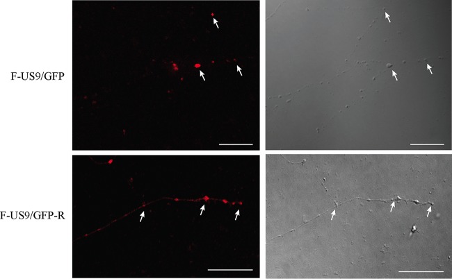
FIG 6.
Confocal micrographs of capsid localization in distal axons in the axonal compartment of neuronal cultures in microfluidic devices infected with US9 deletion (F-US9/GFP) and repair (F-US9/GFP-R) viruses at 22 hpi. Concentration of fluorescence for C capsids (arrows) was visible in varicosities and growth cones in distal axons in in both US9 deletion and repair viruses. Bars, 25 μm.

