FIG 1.
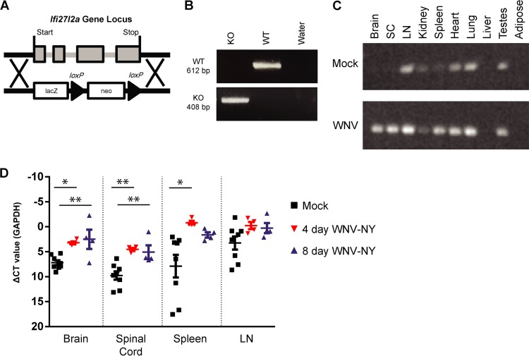
Generation of Ifi27l2a−/− mice and tissue expression of Ifi27l2a. (A) Scheme of Ifi27l2a locus with targeting cassette. Exons are noted in gray, and the target location is noted with a black arrow. Ifi27l2a gene deletion was verified by PCR. Genotyping was verified with a positive-control plasmid containing wild-type Ifi27l2a and negative controls with a null plasmid control. (B) Ifi27l2a deletion was confirmed by the presence of a 408-bp band (KO, knockout), whereas WT Ifi27l2a manifested as a 612-bp band. (C) The Ifi27l2a RT-PCR product was screened for in brain, spinal cord (SC), lymph node (LN), spleen, kidney, lung, liver, white fat (adipose), and testes. Selected mice were infected subcutaneously with WNV-NY, and tissues were collected 4 days after infection and compared to those of mock-infected animals. The results are representative of 2 to 3 mice per treatment group. (D) Following peripheral infection by WNV-NY, selected tissues were collected at 4 and 8 days after infection and expression of Ifi27l2a mRNA was compared to the results seen with mock-infected animals. Ifi27l2a mRNA was measured in brain, spinal cord, spleen, and lymph node and was normalized to the GAPDH gene by qRT-PCR. Means of the results determined for the mock-infected and infected groups were compared using one-way ANOVA followed by Tukey's HSD post hoc analysis (*, P < 0.05; **, P < 0.005). Bars represent means ± standard errors of the means (SEM). CT, threshold cycle.

