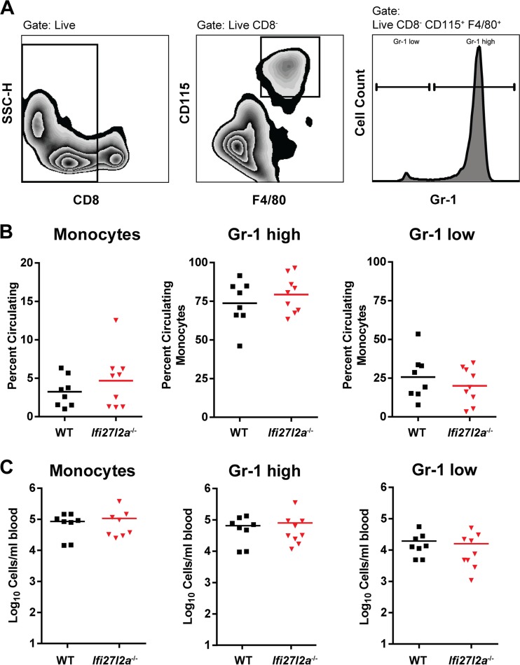
FIG 4.
Circulating monocytes isolated from the blood of WT and Ifi27l2a−/− mice. (A) Circulating blood monocytes were gated as CD8− F4/80+ CD115+ and analyzed for expression of additional surface markers, including Gr-1 (Ly6C and Ly6G). (B and C) WT and Ifi27l2a−/− monocytes were present at similar levels at 8 days postinfection in the blood. Specific monocyte populations of Gr-1high and Gr-1low cells were phenotyped according to prior studies (45, 71), and the results are presented as either percentages (B) or total numbers (C) of cells per milliliter of blood from WT and Ifi27l2a−/− mice. For each group, Student's t test was used to compare cells from WT mice to cells from Ifi27l2a−/− mice. Bars indicate mean values of the results from three independent experiments for 8 to 9 mice for each genotype.

