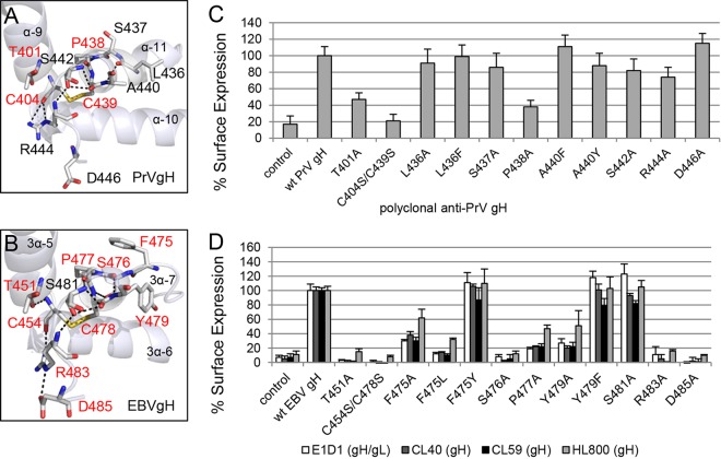
FIG 2.
The DB and contacting amino acids approve the cell surface expression of PrV gH and EBV gH/gL. The arrangements of the conserved amino acids surrounding the conserved DB's connecting three central helices of D-III of PrV (PDB code 2XQY) (A) and EBV gH (PDB code 3PHF) (B) are shown as ribbon diagrams, and the surface expression-required amino acids are highlighted in red. The amino acids (sticks) are colored by element (C and H in gray, N in blue, O in red, and S in orange), and the hydrogen bonds are indicated as black dashes. Surface expression of wt and mutant gH of PrV (C) and EBV (D) is also shown. CELISA was performed with either the monoclonal conformation-specific antibodies against EBV gH (CL40 and CL59) and gH/gL (E1D1) or polyclonal antisera against PrV and EBV gH (HL800). Mean values and standard deviations from three independent experiments are shown.

