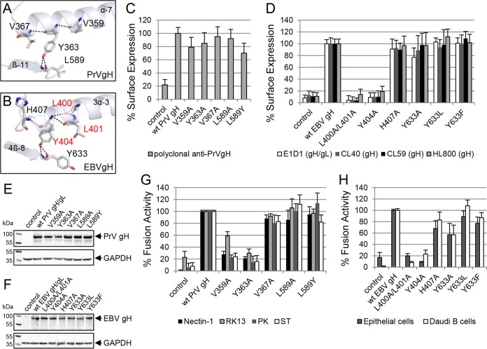
FIG 4.
The conserved tyrosine and a contacting amino acid are important for function of gH during fusion. The conserved tyrosine and contacting amino acids of PrV (PDB code 2XQY) (A) and EBV gH (PDB code 3PHF) (B) are shown as sticks and labeled by element as well as the involved secondary structures are displayed in ribbon diagrams. The surface expression-required amino acids are highlighted in red. Shown are cell surface expression and fusion activities of wt and mutant gH of PrV (C and G) and EBV (D and H). Mean values and standard deviations from three independent experiments are shown. Western blot analysis of wt and mutant gH was performed by using polyclonal antibodies against PrV gH (E) and EBV gH/gL (F) as well as anti-GAPDH.

