
FIG 6.
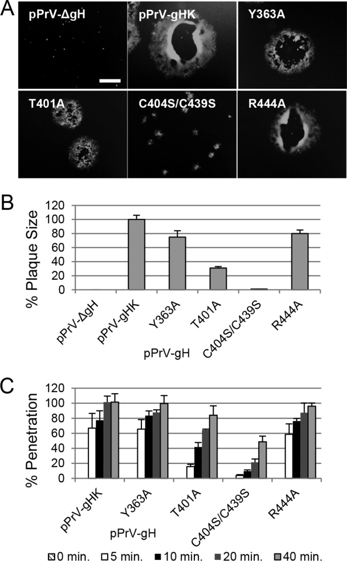
The conserved DB is important for the role of gH during PrV entry. (A) Shown are images of the plaques. Scale bar represents 400 μm. (B) For analysis of the plaque size, 30 plaques were measured in three experiments, with wild-type PrV-Ka set as 100%. (C) For penetration kinetics, RK13 cells were infected on ice for 1 h and incubated for the indicated times at 37°C. Then extracellular virus was inactivated with citric acid. Averages with standard deviations from three independent experiments are shown.