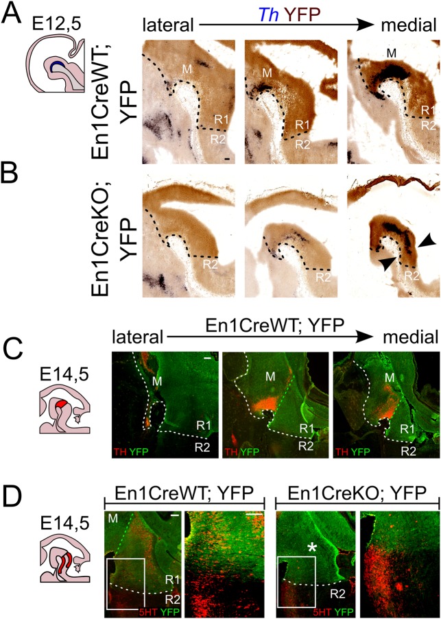
Fig. 3.
Absence of En1 results in changes in the En1-derived R1 area, favoring dopaminergic neurons over serotonergic neurons. (A,C) The region under the control of En1 extends rostrally to the border of dopaminergic neuronal generation in the ventral diencephalon and caudally to the presumed R1/R2 limit at E12.5. The black dotted line delineates the area that is under the control of En1, and therefore YFP-positive. (B) Ectopic DA neurons arise only in an YFP-positive, En1-derived area (arrowheads). (D) Midline section at E14.5 reveals that 5HT is lost in the En1-derived, YFP-area in En1CreKO;YFP (asterisk), but is still present caudal to the R1/R2-boundary. Area of higher magnification indicated by box in D; green dotted line represents the position of the isthmus; the white dotted line delineates the area that is under the control of En1, and therefore YFP-positive. M, midbrain; R1, rhombomere 1; R2, rhombomere 2. Scale bars: 100 μm.

