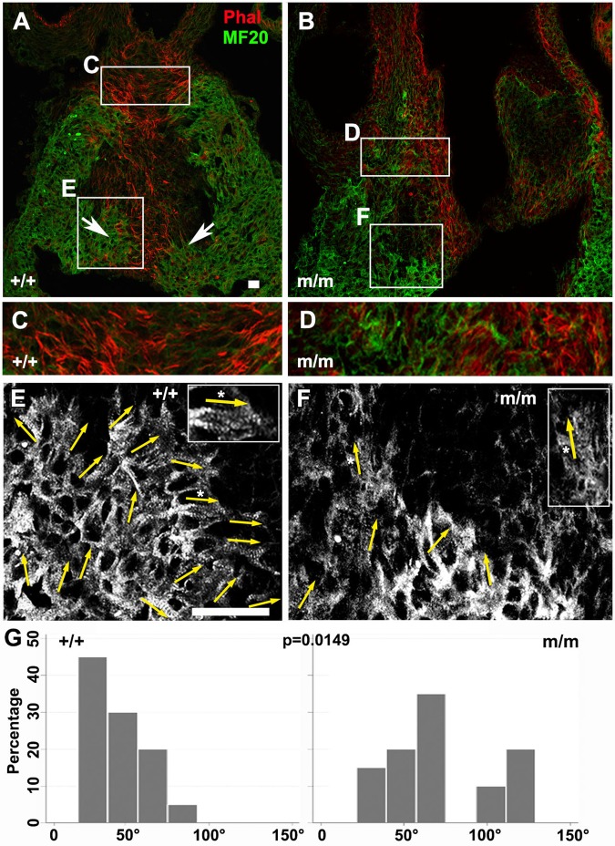
Fig. 5.
Cell polarity and myocardialization defects in the OFT of Bj mutants. (A-F) Phalloidin (red) and MF20 (green) staining of paraffin sections of wild-type (A,C,E,) (n=3) and Bj mutant (B,D,F,) (n=3) heart. In the wild-type heart, phalloidin staining showed actin filament alignment with the direction of cell invasion into the OFT cushion (A,C), but in the Bj mutant, this pattern of actin alignment is not observed (B,D). Arrows in A indicate the two prongs of cardiomyocytes invading the cardiac septum. Examination of the striated banding pattern from the MF20 immunostain showed the developing myofilaments are closely aligned and oriented towards the direction of myocardialization in the wild-type embryo (arrows, E), but in the Bj mutant, the myofilaments are sparse and are largely oriented perpendicular to the direction of myocardialization and septum formation (arrows, F). (G) Quantitation of myofilaments showed wild-type (n=20) cardiomyocytes are polarized in the direction of cell invasion; in Bj mutant hearts (n=20), the cell orientation is not properly aligned (P=0.0149). Quantitative analysis showed cardiomyocytes (MF20 positive) in this conotruncal region of the heart is significantly reduced compared to controls (+/+, +/m 70%; m/m 22%: P=0.0025). Scale bar: 20 µm. The P values were calculated with a two-sample Wilcoxon rank-sum test.

