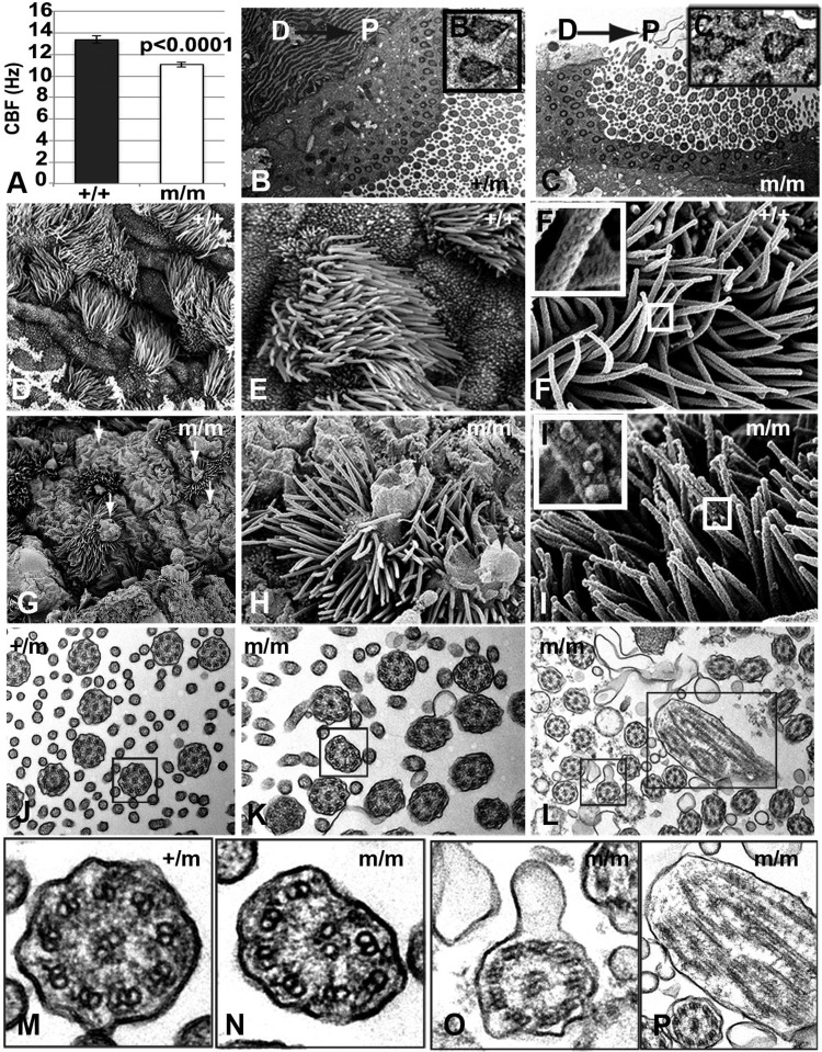
Fig. 8.
Cilia defects in Bj mutant tracheae. (A) Quantitative analysis shows a reduction in cilia beat frequency of Bj mutant tracheal airway cilia. Error bars show standard deviation. (B,C) Transmission electron microscopy (TEM) of motile cilia in both mutant (C) and control (B) trachea show normal planar polarity based on the orientation of basal feet (see boxed insets) on individual cilia towards the proximal (oral) direction. D, dorsal; P, proximal. (D-I). Scanning electron microscopy of Bj mutant trachea (bottom G,H) shows that compared to control (D,E) nonciliated and multiciliated cells have large apical membrane bulges (arrows G,H), which in the multiciliated cells incorporate the axonemes. (I) Some mutant ciliary axonemes also have blebbed surface (boxed I′). (J-L) TEM of Bj mutant cilia show some abnormal axonemal microtubules (K,L), membrane blebs and compound axonemes compared to control (J). (M-P) Enlarged views of (J-L) show the absence of a microtubule doublet in the mutant axoneme (N), blebbed membrane (O), and a compound axoneme (P) compared to the control (M). Compound axonemes are likely contained within the membrane blebs observed by SEM (G,H).

