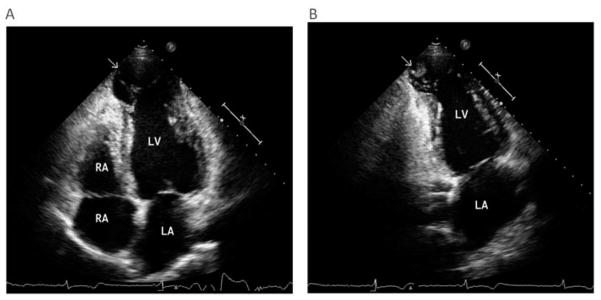
Figure 4. Echocardiography of a patient with chronic chagas cardiomyopathy.
(A) Two-dimensional echocardiography at the apical four-chamber view of the left ventricle demonstrating a dilated apical aneurysm. There is a distinct limit between the normal contractile myocardium and the dyskinetic apex (arrow) with bulging motion during systole. (B) Two-chamber apical view showing an apical thrombus (arrow). There is a mass attached to the left ventricular apical aneurysm with a margin distinct from the underlying wall, which is dyskinetic.
LA: Left atrium, LV: Left ventricle; RA Right atrium; RV Right ventricle.

