Figure 2.
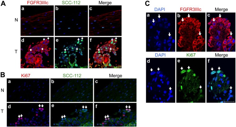
Enhanced expression of FGFR3IIIc was associated with proliferating esophageal carcinoma (EC) cells. (A) Immunofluorescence staining of FGFR3IIIc (a, d) and SCC-112 (b, e) in non-cancerous mucosa (NCM; N, non-tumor) and esophageal squamous cell carcinoma (ESCC) (T, tumor), which was diagnosed as stage 0. In confocal microscopic images, strong staining of FGFR3IIIc (red) was observed in EC cells (d) but not in normal esophageal epithelium cells (a). FGFR3IIIc-positive cells in ESCC were consistent with SCC-112-positive cells (green) (d, e, f: white arrows). Nuclei were stained with DAPI (blue). (B) Immunofluorescence staining of Ki-67 (red; a, d) and SCC-112 (green; b, e) in NCM (N) and ESCC (T). The strong staining of Ki-67 was observed in EC cells, and Ki67-positive cells were consistent with SCC-112-positive cells in ESCC (d, e, f: white arrows). On the other hand, staining of Ki-67 and SCC-112 were not observed in NCM (a, b, c). (C) Immunofluorescence staining of DAPI (a, d), FGFR3IIIc (b), Ki-67 (e) and merged images (c, f) in ESCC samples. The expression of FGFR3IIIc was detected in the same cells, which also expressed Ki-67 in consecutive sections (c, f; white arrows). Scale, 20 μm.

