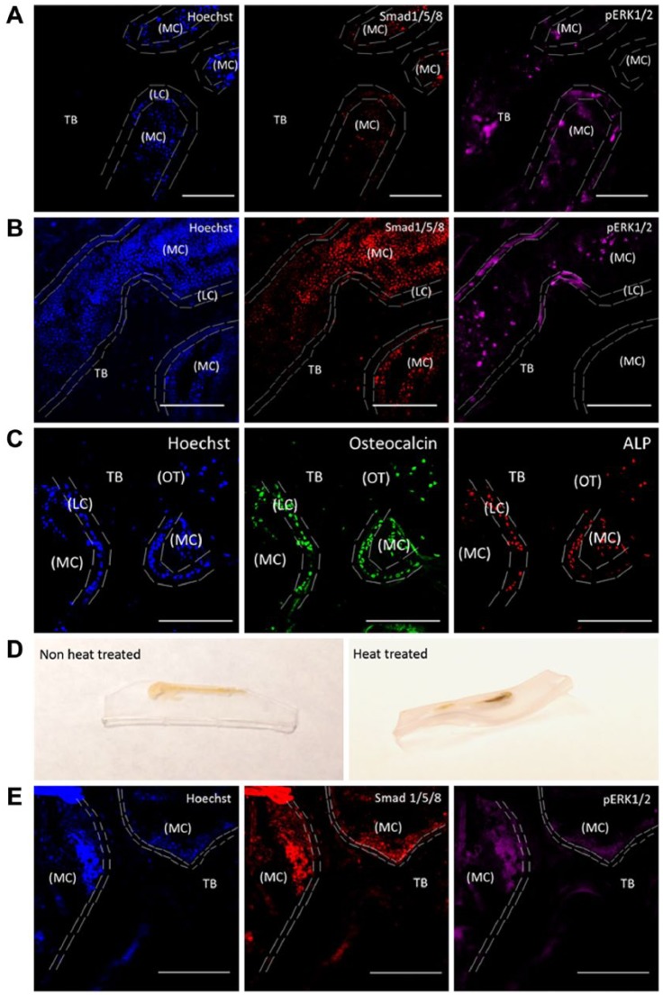
Figure 2.
Testicular hyaluronidase-based antigen retrieval of MMA-embedded femur slices. MMA-embedded samples were immunostained for Smad 1/5/8, pERK1/2, osteocalcin and alkaline phosphatase (ALP). Hoechst staining was used to identify cells within the bone (blue). Heat-induced antigen retrieval was used as a comparison. Sections were imaged in the cancellous or the trabecular bone (TB) of the mouse femur around the marrow cavity (MC) where the lining cells (LC) or the active osteoblasts reside alongside osteocytes (OT). (A) MMA-embedded samples not treated with testicular hyaluronidase and imaged using TPLM. Tissue sections were stained with Hoechst (blue), regions of bone growth and proteins associated with bone cell activity were stained with Smad 1/5/8 (red) and pERK1/2 (magenta). (B) MMA samples treated with testicular hyaluronidase and stained as in (A) were imaged using TPLM. (C) MMA samples treated with testicular hyaluronidase and stained for osteocalcin (green), and ALP (red). (D) Heat-induced antigen retrieval caused morphing of the thick MMA-embedded bone sample if not mounted onto a permanent slide fixture. (E) MMA samples were heat treated and imaged using TPLM. Images were stained with Hoechst (blue), Smad 1/5/8 (red) and pERK1/2 (magenta). All images were taken using 20× magnification. Scale, 100 µM.

