Figure 3.
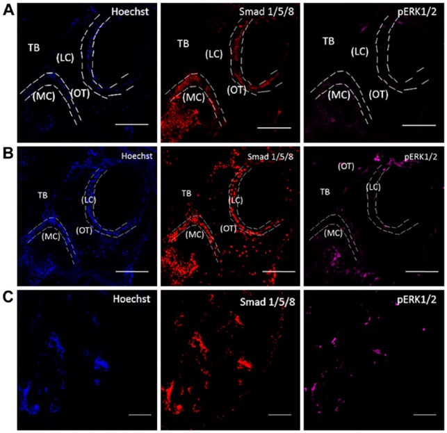
Two-photon excitation laser microscopy imaging of MMA-embedded samples. Imaging of MMA-embedded samples stained with Hoechst (blue), Smad 1/5/8 (red) and pERK1/2 (magenta). (A) Conventional confocal setup. The collected images were of low-resolution and demonstrated high autofluorescence. (B) Two-photon excitation laser microscopy imaging. Images show nuclei and labeling for proteins. (C) A representative tile scan image that was used to examine the entire sample section. Sections were imaged in the cancellous or trabecular bone (TB) of the mouse femur around the marrow cavity (MC) where the lining cells (LC) or active osteoblasts reside alongside osteocytes (OT). Images were taken at 20× magnification. Scale (A, B) 100 µm; (C) 200 µm.

