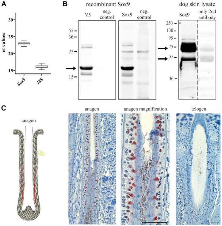
Figure 2.
Sox9. (A) RT-qPCR confirmed the presence of Sox9 mRNA in comparison to 18S rRNA; n=10. (B) Representative western blots. Left panel: Recombinant truncated V5-tagged Sox9 was expressed in E. coli and lysates probed on the same blot with indicated antibodies. The negative control samples were the same as in Fig. 1. Anti-V5 and anti-Sox9 antibodies detected a band corresponding to 20 kDa in size. The calculated size of the recombinant Sox9 protein fragment was 24 kDa. Right panel: Dog skin lysates probed with the same anti-Sox9 antibody revealed two strong bands of 55 kDa and 75 kDa, which were very faint when the anti-Sox9 antibody was omitted. Procedures were as per those specified in Fig. 1. (C) Schematic drawing and immunohistochemical staining for Sox9. Picture on the left: Anagen HF (representative HF for HC stages III-VI) with scattered and discrete positive staining in the innermost layer of the ORS. Picture in the middle: Magnification of the picture on the left. Note the nuclear staining. Picture on the right: Telogen HF. No positive staining was observed. n=5. Note that, for a better overview, the schematic drawings of the hair follicles depict only one follicle of each cycle stage in which a positive staining was observed. Scale (C, left) 50 µm; (C, middle) 25 µm; (C, right) 50 µm.

