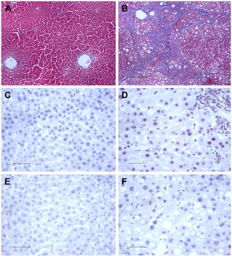
Figure 1.
The histology of hepatic fibrosis. Pathological observations of experimental rat liver sections stained with Masson’s trichrome staining in (A) vehicle control and (B) CCl4-treated liver tissue. The analysis shows that total collagen deposition is significantly increased in CCl4-induced hepatic fibrosis tissues. Compared with the vehicle control (C, E), an increase in positive staining for p-Smad2 (D) and p-Smad3 (F) immunohistochemistry is noted during rat liver fibrosis. Scale, 50 µm.

