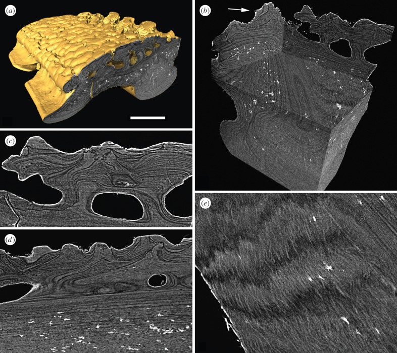
Figure 3.
srXTM histological sections of the dermal skeleton of Vesikulepis funiforma (NHM PV P73708). (a) Transverse section through a body scale; (b) block model constructed from histological slices showing three-dimensional microstructure of the dermal skeleton. The arrow points to a posterior tubercle ridge constructed from discrete, wedge-shaped tubercles; (c,d) transverse sections through the superficial layer and pore canal network of the overlapped area; (e) horizontal section through lamellae of the basal layer showing parallel intrinsic collagen bundles between contiguous lamellae. Scale bar equals 140 µm in (a), 103 µm in (b), 44 µm in (c), 60 µm in (d) and 39 µm in (e).

