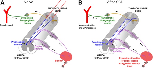
Figure 1.
Schematic illustrating mechanisms involved in the development of autonomic dysreflexia (AD) following SCI.
(A) In intact rats, primary pelvic CGRP+ C-fiber afferents convey sensory information to ascending propriospinal neurons, which relay the transmission from lumbosacral levels to the SPNs in the thoracolumbar spinal cord, eliciting sympathetic response in the peripheral vessels. (B) After high-level SCI, endogenous levels of NGF in the spinal cord are up-regulated and contribute to the plasticity of spinal neuronal pathways, including sprouting of lumbosacral CGRP+ fibers (purple) and propriospinal projections (Lavdas et al., 1997). Thisresults in an exaggerated signal triggered by sensory stimuli, such as distension of the bladder or colon, that is relayed to the reorganized propriospinal projections and then to the uninhibited SPNs. This culminates in a massive sympathetic discharge and baro-reflexic reaction. CGRP: Calcitonin gene-related peptide; SPNs: sympathetic preganglionic neurons; SCI: spinal cord injury.

