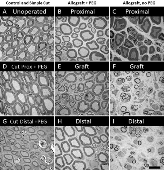
Figure 1.

Typical morphology of plastic embedded, osmium-ferrocyanide stained cross sections of sciatic axons in sciatic nerves that are (A) intact (unoperated), or lesioned 6 weeks previously (B–I).
Lesioned axons are shown: just proximal to (D) or just distal (G) to a single cut that was successfully PEG-fused; just proximal (B), within a 5 mm long allograft (E) or just distal (H) to a successfully PEG-fused allograft; and just proximal (C), within a 5 mm long allograft (F) or just distal to (I) a negative control allograft that was not PEG-fused.
