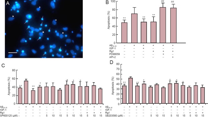
Figure 3.
Rg1 post-treatment protected against Aβ25–35-induced neurotoxicity via ERK1/2 and Akt signaling in hippocampal neurons.
Primary hippocampal neurons were cultured for 24 hours, exposed to Aβ25–35 for 30 minutes, and Rg1 with or without API-2 (10 μM, Akt inhibitor) or PD98059 (10 μM, MEK inhibitor), SP600125 (5, 10 or 15 μM, p38 MAPK inhibitor), or SB203580 (5, 10 or 15 μM, JNK inhibitor) was added for another 48 hours. Insulin-like growth factor-1 (IGF-1) was added to the culture as a positive control. Normal controls were not exposed to Aβ25–35. (A) Hoechst 33258 staining: apoptotic (arrows) and viable (arrowheads) cells. (B–D) Apoptosis of hippocampal neurons in Rg1 with API-2 or PD98059 (B), SP600125 (C) or SB203580 (D). Data are the mean ± SEM of five individual experiments. *P < 0.05, **P < 0.01, vs. Aβ25–35; ##P < 0.01, vs. Aβ25–35 + Rg1 (one-way analysis of variance followed by Newman-Keuls post hoc test). Scale bar in A: 200 μm.

