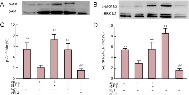
Figure 4.
Rg1 post-treatment reversed Aβ25–35-induced reduction of ERK1/2 and Akt phosphorylation in hippocampal neurons.
Primary hippocampal neurons were cultured for 24 hours, exposed to 20 μM Aβ25–35 for 30 minutes, and Rg1 with or without 10 μM API-2 was added for another 48 hours. In addition, insulin-like growth factor-1 (IGF-1) was added to the culture as a positive control. Normal control cells were not exposed to Aβ25–35. (A, B) Western blot analysis of total (t-Akt) and phosphorylated Akt (p-Akt) (A) and total (t-ERK) and phosphorated ERK (p-ERK). (C, D) Phosphorylation levels of Akt (C) and ERK (D). Data are the mean ± SEM of three individual experiments. **P < 0.01, vs. Aβ25–35; ##P < 0.01, vs. Rg1 + Aβ25–35 (oneway analysis of variance followed by Newman-Keuls post hoc test).

