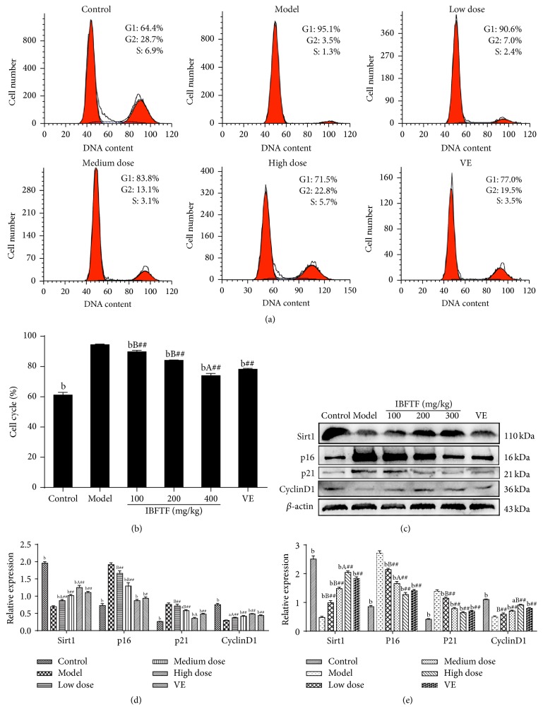
Figure 2.
The IBFTF influences the expression of Sirt1 to indirect regulation of p16INK4a and P53-P21Cip1 pathways. The cell cycle distribution of the cells was analyzed by flow cytometry. The representative graphs are shown in (a). The quantitative analysis is demonstrated as histograms in (b). The protein levels of regulators of cell cycle were detected by western blot in (c) and (d). (e) The mRNA levels of regulators of cell cycle were detected by RT-qPCR. The data are presented as means ± SD (n = 4) and all experiments were done in triplicate. a P < 0.05; b P < 0.01 compared with model group (all groups). A P < 0.05; B P < 0.01 compared with Vit E group (IBFTF groups). # P < 0.05; ## P < 0.01 compared with control group (Vit E and IBFTF groups).

