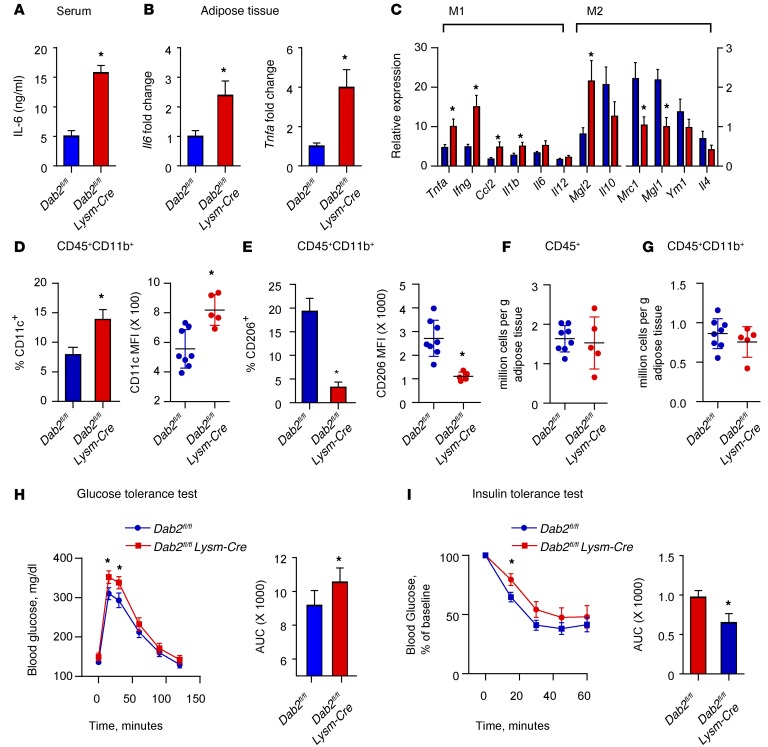
Figure 5. DAB2 in myeloid cells regulates HFD-induced adipose tissue inflammation and insulin resistance.
Dab2fl/fl and Dab2fl/fl Lysm-Cre female mice were fed an HFD for 12 weeks. (A) Serum IL-6 levels after an HFD for 12 weeks as analyzed by ELISA. (B) Il6 and Tnfa mRNA levels in adipose tissue were analyzed by qPCR. (C) pPCR phenotypic characterization of cells isolated from the SVF in adipose tissue. (D–G) Cells isolated from the SVF in gonadal adipose tissue of HFD-fed Dab2fl/fl and Dab2fl/fl Lysm-Cre mice were stained with CD45-APC-Cy7, CD11b-FITC, CD206-PE, and CD11c-AF647 Abs and subjected to FACS analysis. CD45+CD11b+ cells were analyzed for the percentage of cells that were CD11c+ and for the MFI of CD11c (D), and CD45+CD11b+ cells were analyzed for the percentage of cells that were CD206+ and for the MFI of CD206 (E). Each point represents 1 mouse in the MFI graphs in D and E. *P = 0.003, by 2-tailed, unpaired Student’s t test (D) and *P = 0.004, by 2-tailed, unpaired Student’s t test with Welch’s correction (E). Absolute numbers of CD45+ (F) and CD45+CD11b+ (G) cells per gram of gonadal adipose tissue were quantified and showed no difference in cell infiltration levels between HFD-fed Dab2fl/fl and Dab2fl/fl Lysm-Cre mice. For glucose tolerance tests (H) and insulin tolerance tests (I), mice were fasted for 6 hours and injected i.p. with a 1 g/kg glucose bolus or 0.75 U/kg insulin, respectively, and blood glucose levels were measured in tail blood by glucometer. Data were analyzed by calculating the AUC and represent the mean ± SEM. n = 10–12. *P < 0.05, by 2-tailed, unpaired Student’s t test.

