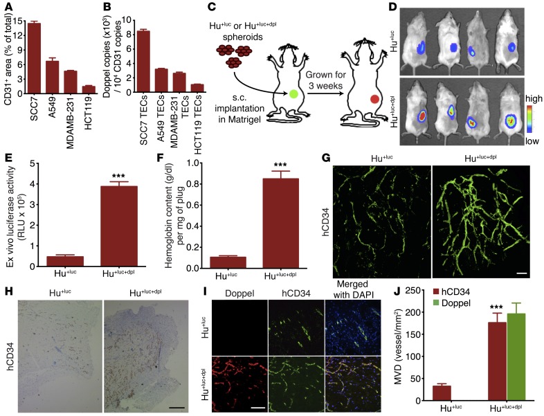
Figure 2. Increased doppel expression increases tumoral angiogenesis and EC function.
(A) Total volume of blood vessels in squamous, lung, breast, and colon tumor. An aliquot of tumor (1 g) was dissected to make single cells from the site where the CD31-positive area was calculated (n = 3 sections from each tumor). (B) Doppel expression in individual TECs as determined by flow cytometric analysis (3 experiments). (C) Experimental procedure for evaluation of the gain-of-function effect of doppel in ECs. Luciferase-expressing HUVECs (Hu+luc) and doppel-transfected Hu+luc (Hu+luc+dpl) spheroids were implanted s.c. in a Matrigel-fibrin matrix into female SCID mice. Three weeks after transplantation, the vascularization was analyzed. (D) Noninvasive monitoring of vascularization by bioluminescence imaging (n = 4 mice). (E) Ex vivo bioluminescence counts. ***P < 0.001 versus Hu+luc, Student’s t test. (F) Hemoglobin content within Hu+luc and Hu+luc+dpl plugs was quantified. ***P < 0.001 versus Hu+luc, Student’s t test. (G) 3D structure of the vascular network formed by Hu+luc and Hu+luc+dpl cells, as assessed by confocal microscopy using IF whole-mount staining for hCD34. Scale bar: 50 μm. (H) Immunoperoxidase detection of hCD34-positive blood vessels in Hu+luc and Hu+luc+dpl plugs. Scale bar: 20 μm. (I) Characterization and images of vascular network by staining for doppel (red), hCD34 (green), and nuclei (blue) in Hu+luc and Hu+luc+dpl plugs. Scale bar: 20 μm. (J) Quantification of hCD34-positive and doppel-positive mean vessel density (MVD) in Hu+luc and Hu+luc+dpl plugs. Doppel-positive vessels were not detected in Hu+luc plugs. ***P < 0.001 versus Hu+luc, Student’s t test. n = 4 plugs per experiment.

