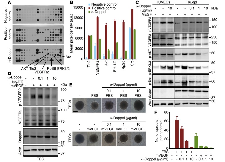
Figure 3. Doppel plays a role in VEGFR2 signaling.
(A) Phosphorylated RTK (p-RTK) signaling array of Hu.dpl exposed to fasting media, complete media, and α-doppel (30 minutes, 10 μg/ml) in the presence of complete media. (B) Quantification of pixel density of p-Tie2, p-VEGFR2, p-AKT, p-ERK1/2, p-RpS6, and p-Src. See also Supplemental Figure 10. (C) Immunoblots of p-VEGFR2, p-AKT, p-ERK1/2, p-Src, and total VEGFR2, doppel, and actin in HUVECs and Hu.dpl cells treated with different concentrations of α-doppel in the presence of VEGF165 (100 ng/ml). Cells were pretreated with α-doppel for 30 minutes, and then VEGF165 was added for 5 minutes. (D) Immunoblots of p-VEGFR2, total VEGFR2, total doppel, and actin in TECs treated with different concentrations of α-doppel in the presence of mVEGF (100 ng/ml). Dose-dependent inhibition (E) and total number of TEC sprouts (F) by α-doppel stimulated with either 10% FBS or mVEGF (100 ng/ml). Scale bar: 100 μm. Each experiment was repeated 3 times.

