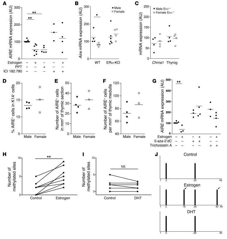
Figure 5. Pathways involved in estrogen-induced downmodulation of thymic AIRE expression.
In vitro effects of the estrogen agonist PPT (10 nM) and the ER antagonist ICI 182.780 (20 μM) on AIRE mRNA expression in TECs (n = 3–7) (A). Aire (B) and AIRE-dependent TSA (Chrna1, and thyroglobulin) (C) mRNA expression in male and female thymuses of ERα-KO mice (n = 4–6). Percentage of cells expressing the AIRE protein among K14+ cells (D), number of AIRE-expressing cells per mm2 of thymic section (E) and per mm2 of thymic medulla (F), of male and female ERα-KO mice (n = 3–4). Effects of 5-aza-2′-deoxycytidine (5-aza-2′dC) (5 μM), a DNA methylation inhibitor, and trichostatin A (50 nM), a histone deacetylase inhibitor on AIRE mRNA expression in TEC cultures (G) (n = 3–4). Effect of estrogen (1 nM) (H) and DHT (1 nM) (I) on methylation site levels of CpG motifs in the human AIRE promoter in independent cultured human TECs (8 for estrogen and 6 for DHT). Representative methylation pattern of the CpG motifs within the human AIRE gene promoter in primary cultured TECs treated with 1 nM of estrogen or of DHT (J). Each bar represents the methylation of a single CpG motif. The arrows show methylation sites that appeared after estrogen treatment. P values were obtained using the 1-way ANOVA test (A and G), the Mann-Whitney U test for unpaired data (B–F), and the Wilcoxon test for paired data (H and I). *P < 0.05 to 0.01; **P < 0.001.

