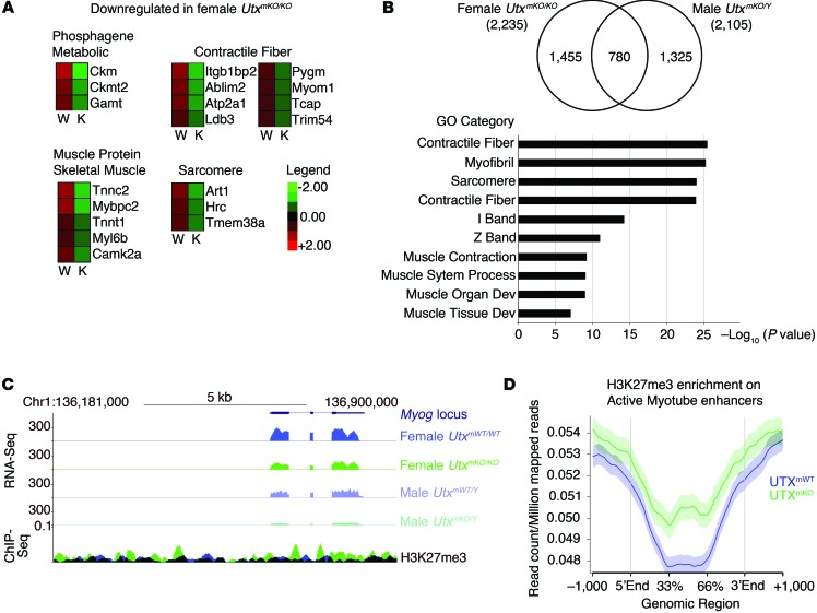
Figure 4. UTX regulates the muscle gene expression program through demethylation of H3K27me3 marks.
WT primary myoblasts were isolated from female or male UtxmKO/TdT or UtxmWT/TdT mice. After expansion in culture for 5 days, cells were treated with 4-OH-tamoxifen for 24 hours. Cells that underwent recombination were isolated by FACS, plated, and allowed to adhere to the plate overnight. Myogenic differentiation was then induced for 24 hours prior to RNA isolation. (A and B) RNA-Seq analysis was performed to identify genes whose expression is modified in differentiation myoblasts from UtxmKO/TdT compared with UtxmWT/TdT mice. (A) A heatmap is shown for selected genes of different ontologies that are differentially expressed (P < 0.05). (B) The overlap of 780 genes that are downregulated in both male and female UtxmKO/TdT mice is represented by a Venn diagram. GO analysis of the 780 genes downregulated in myoblasts from both males and female UtxmKO/TdT shows highly significant enrichment of genes involved in muscle development and function. (C and D) H3K27me3 ChIP-Seq analysis was performed to examine H3K27me3 enrichment in differentiating myoblasts isolated from UtxmKO/TdT (green) or UtxmWT/TdT (blue) mice. (C) RNA-Seq and H3K27me3 ChIP-Seq tracks mapping to the Myog locus are presented in UtxmKO/TdT or UtxmWT/TdT conditions. (D) Analysis of H3K27me3 levels at enhancers that have previously been established as myotube-specific enhancers (39).

