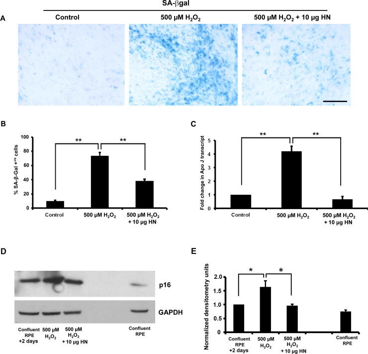
Figure 11.
Humanin delays oxidative stress–induced senescence in RPE cells. The RPE cells were treated with 500 μM H2O2 or 500 μM H2O2 plus 10 μg/mL HN and processed for SA-β-Gal staining or p16INK4a immunoblot. Oxidative stress significantly increased the number of SA-β-Gal–positive cells and RPE cell senescence was considerably lower with HN cotreatment (A, B) The H2O2 treatment induced 75% cell senescence, whereas the HN cotreatment showed less than 30% cell senescence (B). (C) Gene expression of senescence marker gene ApoJ by real-time PCR (forward, 5′-AGAGTGTAAGCCCTG CCTGA-3′; reverse, 5′-CATCCAGAAGTAGAAGGGCG-3′). The expression of ApoJ was significantly higher in H2O2-treated cells than in control RPE cells. The increase in expression of ApoJ was significantly suppressed when cells were coincubated with HN. (D, E) Oxidative stress significantly increased expression of p16INK4a, while HN cotreatment restored p16INK4a expression to that of control cells. Expression of p16INK4a was considerably low in normal confluent RPE cells. *P < 0.05, **P < 0.01.

