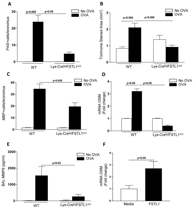
Figure 3. Inhibition of macrophage Fstl1 expression inhibits airway remodeling.
Lys-Cretg/Fstl1Δ/Δ or WT mice (8 mice/group) were sensitized with OVA allergen followed by chronic exposure to OVA allergen. Levels of lung mucus were quantitated by PAS staining (Fig. 3a). Levels of peribronchial trichrome staining were quantitated by image analysis (Fig 3b). The number of peribronchial eosinophils was quantitated by MBP immunostaining and image analysis (Fig. 3c). Levels of oncostain M (OSM) were quantitated by qPCR (Fig. 3d). Levels of BAL MMP9 were quantitated by ELISA (Fig. 3e). In separate experiments, WT mouse bone marrow derived macrophages were incubated for 24 hrs with either Fstl1 (100 ng/ml) or media and levels of OSM mRNA quantitated by qPCR (Fig. 3f).

