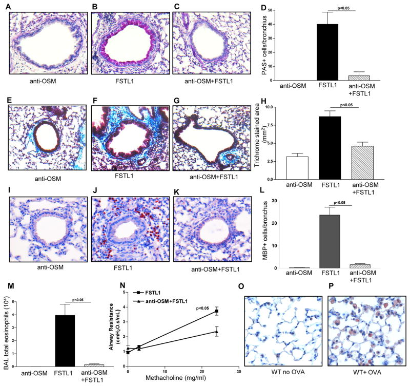
Figure 5. Blocking Oncostain M inhibits Fstl1 induced airway remodeling.
WT mice (4 mice/group) were administered Fstl1 intranasally daily for 15 days, with or without pre-treatment with an anti-oncostatin M antibody (anti-OSM). A control WT group received the anti-oncostatin M antibody and no Fstl1. Levels of lung mucus were quantitated by PAS staining (Fig. 5a–d). Levels of peribronchial trichrome staining were quantitated by image analysis (Fig. 5e–h). The number of MBP+ peribronchial eosinophils were quantitated by image analysis (Fig. 5i–l). The number of Wright-Giemsa stained BAL eosinophils was quantitated by light microscopy (Fig. 5m). Levels of airway responsiveness to methacholine was assessed by flexivent (Fig. 5n). In a separate experiment, lungs from either WT mice subjected to chronic OVA challenge (WT+OVA), or WT mice not challenged with OVA (WT+ No OVA), were immunostained with an anti-OSM Ab to detect OSM positive cells in the lung.

