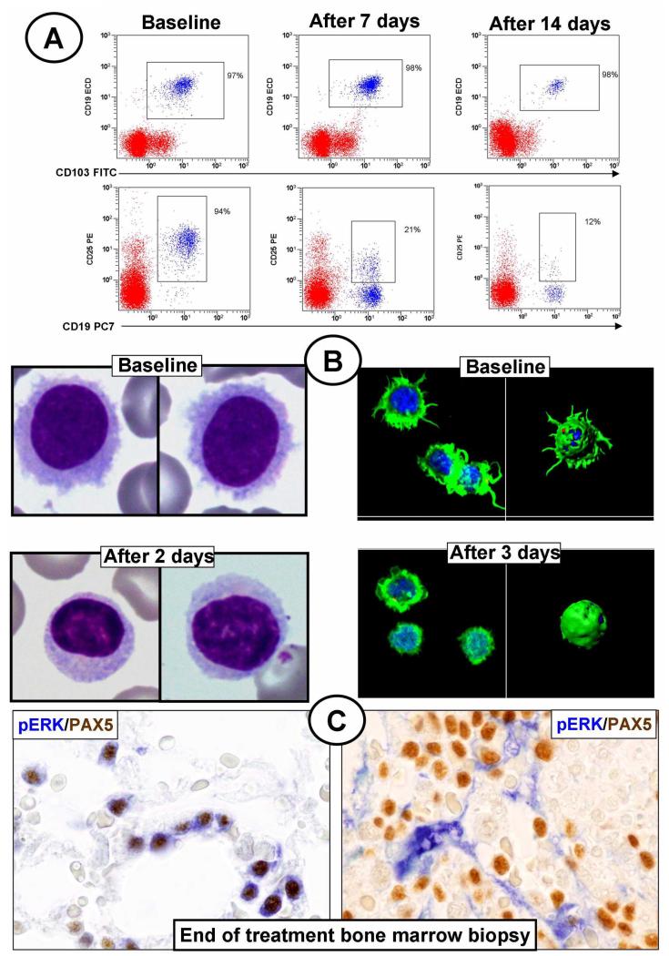
Figure 3. Phenotypic and molecular changes of HCL cells during treatment with vemurafenib in the Italian trial.
(A) Flow cytometry dot plots of a patient’s BM aspirate gated on CD45 cells and showing at baseline (left panels) CD25 expression by 94% of leukemic cells (CD19+/CD103+, upper row; CD19+/CD25+, bottom row). A progressively loss of CD25 expression (but not of CD19 or CD103 expression) is seen over 1 and 2 weeks of treatment with vemurafenib (down to 21% and 12%, respectively; middle and right panels, respectively). The remaining cells (red events) are normal CD45+ hematopoietic cells. (B) Left panels. May-Grümwald-Giemsa staining of a patient’s peripheral blood smear featuring leukemic cells rich in hairy projections at baseline (top), but not after 2 days of vemurafenib treatment (bottom). Right panels. Confocal fluorescence microscopy analysis of blood leukemic cells purified from the same patient shows prominent surface projections (stained in green by phalloidin) at baseline (top), but not after 3 days of vemurafenib treatment (bottom); these changes are clearly evident both in electronically magnified two-dimensional and three-dimensional reconstructed images of representative cells; (C) Double immunostaining of BM biopsy taken the day after the end of treatment and double stained for PAX5 (a B-cell marker; in brown) and phospho-ERK (pERK; in blue). In some responding patients (one exemplified in the left panel) persistence of pERK+ leukemic hairy cells is observed; conversely, in other patients (one exemplified in the right panel) residual hairy cells do not detectably express phospho-ERK, with stromal cells strongly positive for phospho-ERK as internal positive control.

