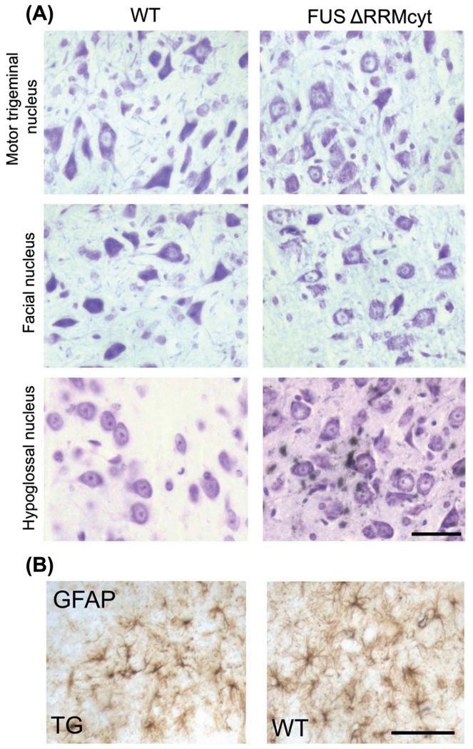
Figure 4. Brainstem motor nuclei of FUS ΔRRMcyt mice do not display obvious signs of neurodegeneration.
(A) Motor trigeminal, facial and hypoglossal nuclei in TG and WT mice were stained with cresyl violet to permit identification of motor neurons. No morphological differences were identified in TG mice compared with WT littermates. (B) Immunoreactivity to glial fibrillary acidic protein (GFAP) and morphology of astrocytes are indistinguishable between FUS ΔRRMcyt mice and WT littermates. Representative images shown. Scale bars: A, 25 μm; B, 50 μm.

