Figure 3.

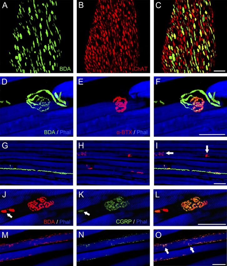
Three-dimensional projections from stacks of CLSM images showing BDA tracer combined with fluorescence staining of other cellular molecules. A–C, Tracer visualization with avidin (green) was combined with anti-ChAT labeling (red). A, Tracer-positive axons at the entry site of the abducens nerve in the lateral rectus muscle. B, The axons of this abducens nerve exhibit ChAT immunoreactivity. C, Overlay of A and B in yellow mixed color showing that all tracer-positive axons also exhibit ChAT immunoreactivity, but other ChAT-positive axons contained no tracer. D–I, Triple fluorescence staining with streptavidin (green), α-bungarotoxin (red), and phalloidin (blue) of a medial rectus muscle. D, G, Tracer-positive axons establishing neuromuscular contacts resembling en plaque motor terminals (D) and en grappe motor terminals (G). E, H, En plaque and en grappe motor terminals, respectively, after α-bungarotoxin labeling. F, I, Overlays in yellow mixed color confirming that tracer-positive en plaque-like and en grape-like endings bind α-bungarotoxin. Other motor terminals (arrows) lack tracer (I). J–O, Triple fluorescence staining with streptavidin (red), anti-CGRP (green), and phalloidin (blue) J, M, Tracer-positive axons establishing en plaque and en grappe motor terminals, respectively. K, N, Tracer-positive axons and motor terminals exhibit CGRP immunoreactivity. The arrows in J and K show the tracer (BDA)-positive axon (in J), which also showed CGRP immunolabeling (in K). L, O, Overlays in yellow mixed color. En grappe endings are indicated by arrows (O). Scale bars, 50 μm.
