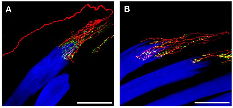
Fig. 1. CLSM images of palisade endings. Nerve fibers are labeled with anti-neurofilament (red), nerve terminals with anti-synaptophysin (green), and muscle fibers with phalloidin (blue). Nerve terminals which exhibit neurofilament and synaptophysin immunoreactivity appear yellow in the overly. The tendon not labeled continues the muscle fiber to the right.
A. A neurofilament positive nerve fiber runs along the muscle fibers into the tendon, forms a 180° turn and ends in an arborization at the muscle fiber tip. The nerve terminals of the palisade ending are synaptophysin positive and are located in the tendon and around the muscle fiber tip. B. Showing another palisade ending. Only the peripheral part of the nerve fiber supplying the palisade ending is shown. Palisade nerve terminals are synaptophysin positive. Scale bars, 100 μm.

