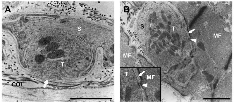
Fig. 4.
TEM micrographs of palisade endings. A. High resolution micrograph of a neurotendinous contact in a palisade ending. The nerve terminal (T) contains mitochondria and numerous clear vesicles and is partly covered by a Schwann cell (S). At the contact point only a basal lamina (arrow) separates the nerve terminal and collagen (COL). B. High resolution micrograph of a neuromuscular contact in a palisade ending. A Schwann cell covers part of the nerve terminal which contains mitochondria and clear vesicles. The basal lamina (arrow) between muscle fiber and nerve terminal is in some parts absent (arrowhead). A detail of the nerve terminal is shown in the inset. Scale bars, 1 μm.

