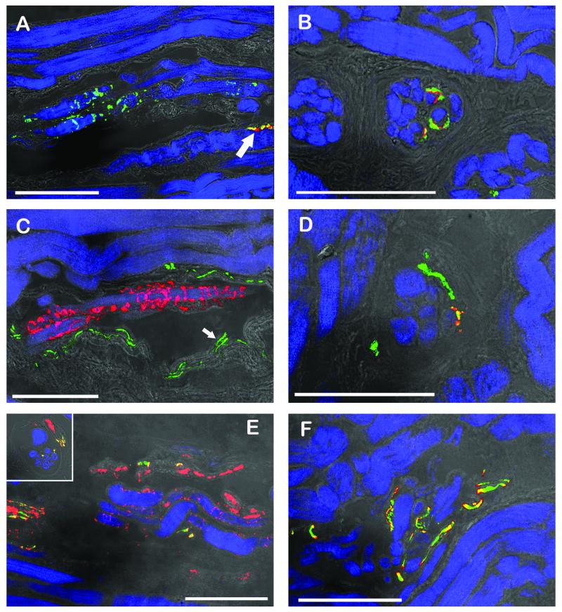
Fig. 3.
CLSM images of muscle spindles showing the equatorial regions (A, C, E) and the polar regions (B, D, F). The equatorial regions are shown in longitudinal sections whereas the polar regions are shown in cross section (B, D) or oblique section (F) respectively. Triple fluorescent images were overlaid with a transmission light image. (A, B) Labeling with anti-synaptophysin (green), α-bungarotoxin (red) and phalloidin (blue). (A) Sensory nerve endings in the equatorial region are only positive for synaptophysin whereas a motor terminal (arrow) outside the spindle is synaptophysin/α-bungarotoxin-positive. (B) In the muscle spindle’s polar region, motor terminals exhibit synaptophysin/α-bungarotoxin reactivity. (C, D) Labeling with anti-ChAT (green), anti-synaptophysin (red), and phalloidin (blue). (C) Sensory anulospiral nerve endings in the equatorial regions are synaptophysin-positive but ChAT-negative. ChAT-positive nerve fibers are seen alongside the spindle. Two ChAT-positive axons (arrow) running towards the spindle’s pole are visible inside the spindle. (D) In the polar region ChAT-positive nerve fibers establish motor terminals on the intrafusal muscle fibers. Motor terminals co-localize ChAT and synaptophysin. (E, F) Labeling with anti-ChAT (green), anti-neurofilament (red), and phalloidin (blue). (E) In the muscle spindles’ equatorial region nerve fibers solely positive for neurofilament and nerve fibers positive for ChAT/neurofilament are visible. Only neurofilament-positive axons enwrap intrafusal muscle fibers and are thereby forming anulospiral endings in the equatorial region. Inset showing the mixed innervation of a muscle spindle in a cross section. (F) In the polar region axons stain double positive for ChAT and neurofilament. Scale bars, 100 μm.

