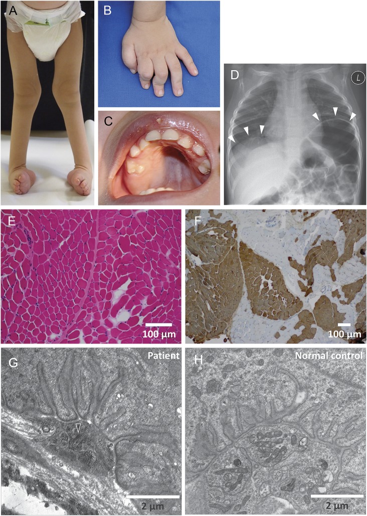
Figure 1. Clinical images of the patient.
(A) Talipes equinovarus foot deformity. (B) Arthrogryposis of the hand, with inability to extend the fingers. (C) High-arched palate as a sign of intrauterine muscle weakness. (D) Diaphragmatic palsy with eventration (white arrowheads). (E) Hematoxylin and eosin staining of the quadriceps muscle rules out grouped fiber atrophy. (F) Staining with anti-myosin heavy chain (fast) antibodies reveals fiber type grouping as a sign of denervation/reinnervation. Type II fibers are depicted in brown. (G) Electron microscopy of the neuromuscular endplate reveals condensation of the axoplasm and degeneration of axonal organelles (open arrowhead), including mitochondria and loss of presynaptic neurofilaments, while the subneural clefts appear normal. Five of 5 endplates that could be discovered in the muscle biopsy specimen had identical abnormalities. (H) Normal neuromuscular endplate of the quadriceps muscle of an age-matched control at the same magnification for comparison.

