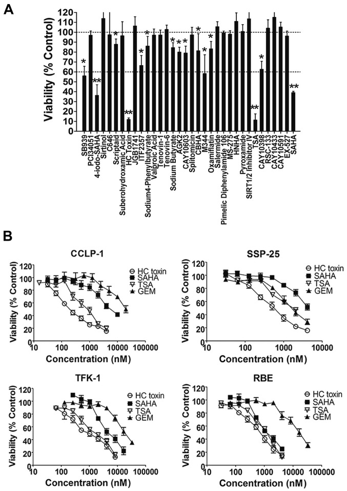
Figure 1.
Results of the HDAC inhibitor screening in intrahepatic cholangiocarcinoma (ICC) cell lines by cell viability assays. (A) Cell viability of RBE cells after being co-cultured with 34 HDAC inhibitors for 48 h. All of the HDAC inhibitors were used at a final concentration of 3 µM. Differences were statistical significant at *P<0.05 compared with 100% and **P<0.05 compared with 60% viability. (B) Cell viability of four ICC cell lines after being co-cultured with HC toxin, SAHA, TSA or gemcitabine (GEM) for 48 h. The concentration gradient used increased from 30 nM to 30 µM with a common ratio of two.

