Figure 5.
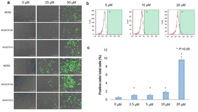
PRIMA-1MET induces intracellular ROS accumulation in EOC cells. (a) Intracellular ROS accumulation after 16 h of PRIMA-1MET treatment in the NOS2 and NOS3 cells, and their chemo-resistant cells. Intracellular ROS levels were detected by CM-H2DCFDA, resulting in fluorescence positivity under fluorescence microscopy. (b) Dose-dependent intracellular ROS accumulation in TOV21G cells. TOV21G cells were maintained in medium with the indicated concentrations of PRIMA-1MET, labeled with 5 µM CM-H2DCFDA, and then subjected to flow cytometry. The percentage of cells with fluorescence intensity above the level of 1,000 FL was measured. (c) Bars represent the mean percentage of fluorescence-positive cells. Error bars represent standard deviations.

