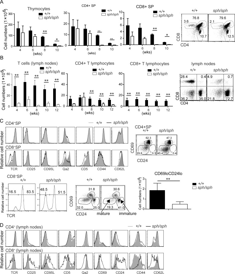
Fig 1. Gimap5 deficiency results in disrupted T cell development.
(A) Total number of thymocytes from wildtype and Gimap5sph/sph mice at different ages (in weeks) was counted by trypan blue staining (bar graphs). Flow cytometric analysis of thymocytes following staining with anti-CD4 and anti-CD8 antibodies in wild-type and Gimap5sph/sph mice. (B) Total number of T, CD4+ and CD8+ T cells in pooled lymph nodes (inguinal, axillary, brachial and superficial cervical) from wildtype and Gimap5sph/sph mice at different ages (in weeks, from the same groups of mice as in A) were counted by trypan blue staining. The absolute numbers of total T, CD4+ and CD8+ T cells were calculated by factoring the frequency of CD3+ T cells and the frequency of CD4+ and CD8+ cells within the CD3+ gate. (C) The phenotype of CD4+ or CD8+ SP thymocytes from wild-type and Gimap5sph/sph mice for the indicated markers is shown as histograms. The data for TCR expression shown in the enlarged histogram and the overlap histogram for CD8+SP are from different mice. (D) The phenotype of CD4+ or CD8+ T cells from wild-type and Gimap5sph/sph mice for the indicated markers is shown as histograms. Data shown are representative of 3 independent experiments. Each experiment consisted of analyzing 2 individual sex- and age-matched mice from each genotype.

