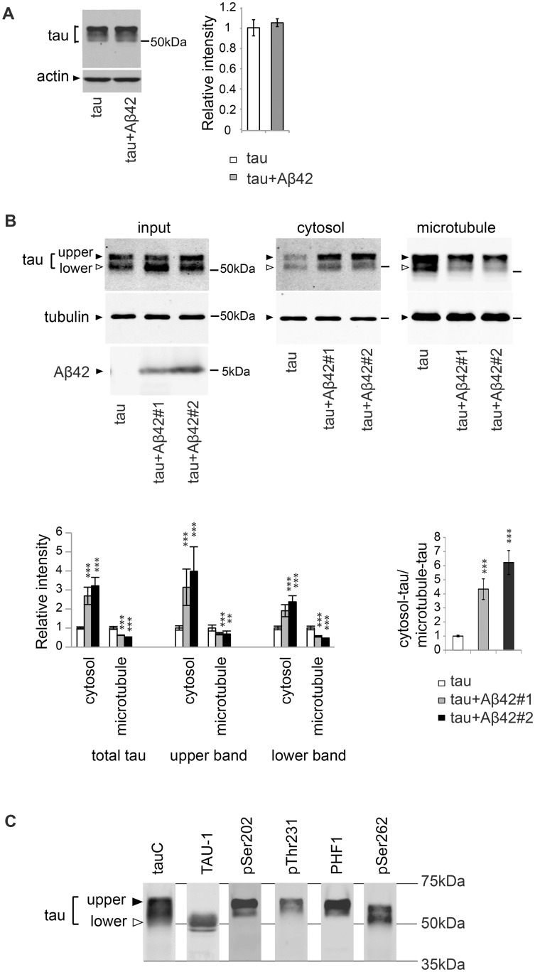
Fig 1. Aβ42 increases the level of tau in the cytosol and decreases the level of microtubule-bound tau.
(A) Aβ42 does not change total tau levels. Western blot of heads of flies expressing tau alone (tau) or that co-expressing tau and Aβ42 (tau+Aβ42) driven by gmr-GAL4 with anti-tau antibody. Actin was used as a loading control. Mean ± SD, n = 5; no significant difference was found by Student's t-test (p>0.05). Representative blots are shown. (B) Co-expression of Aβ42 increases the levels of tau free from microtubules and reduces the levels of tau bound to microtubules. The levels of tau and tubulin in the lysate of fly heads expressing tau alone (tau) or co-expressing tau and Aβ42 (tau+Aβ42#1 and tau+Aβ42#2) before sedimentation (input), in the supernatant (cytosol) and in the pellet containing microtubules (microtubule) were analyzed by western blotting by using anti-tau antibody. The same amount of proteins from each genotype was loaded. Expression of Aβ42 was confirmed by western blot with anti-Aβ antibody (Aβ42). Two independent transgenic fly lines expressing Aβ42 at different expression levels (Aβ42#1 and Aβ42#2) yielded similar results, and the fly line with higher Aβ42 (Aβ42#2) expression exhibited a larger effect. Transgene expression was driven by gmr-GAL4. Mean ± SD, n = 4; **, p<0.01, ***, p<0.005 compared to tau by one-way ANOVA with Tukey's post hoc test. Representative blots are shown. (C) Two major bands detected by western blotting of fly heads expressing tau with a pan-tau antibody (tauC) differ in their phosphorylation patterns. Western blots with TAU-1 antibody, which specifically recognizes tau protein without phosphorylation at several AD-related sites (Ser194, Ser195, Ser198, and Ser202) (TAU-1), anti-phospho-Ser202, anti-phospho-Thr231, anti-phospho-Ser396/404 (PHF1), or anti-phospho-Ser262 antibody (pSer262) are shown. Transgene expression was driven by gmr-GAL4.

