
Figure 4. Down-regulation of p55PIK re-sensitized GIST882IR tumors to Imatinib in vivo.
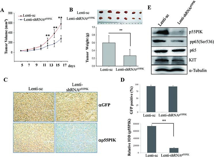
GIST882IR cells were resuspended in culture medium (2 × 10⁷ cells/ml) and injected subcutaneously into athymic nude mice (100 μl/tumor). At day 7 after inoculation Imatinib was given by intragastric administration (100 mg/kg) daily as well as Lenti-sc or Lenti-shRNAp55PIK (1010 tu/20 μl) was injected into tumors every 3 days. A. Down-regulation of p55PIK decreased the tumor volume of GIST882IR in vivo. Tumor volume was measured every 2 days. *p < 0.05; **p < 0.01. B. Down-regulation of p55PIK potentiated the inhibitory effects of Imatinib on the tumor growth of GIST882IR cells in vivo. Tumors injected with Lenti-sc or Lenti-shRNAp55PIK were weighed and analyzed. C. Immuno-histochemical (IHC) analysis of tumor samples using anti-GFP and anti-p55PIK antibodies. Representative images from IHC analysis detecting GFP signal (upper panel) or p55PIK signal (lower panel) shown. D. Upper panel: the percentage of GFP-positive cells in tumor samples was determined and used as an indicator to show the lentivirus transfection efficiency from intratumoral injection (mean ± SE (n = 3)). Lower panel: IHC quantification of p55PIK expression. The expression of p55PIK in Lenti-sc or Lenti-shRNAp55PIK-treated tumor samples were examined by IHC and the intensity of signal (Relative IOD) was determined and analyzed. **p < 0.01. E. Down-regulation of p55PIK decreased the expression of KIT and phosphorylation of NF-κB p65 in xenograft tumors. The expression of p55PIK, p65, pp65(Ser536) and KIT in tumors from GIST882IR cells injected with Lenti-sc or Lenti-shRNAp55PIK was examined by Western blotting.