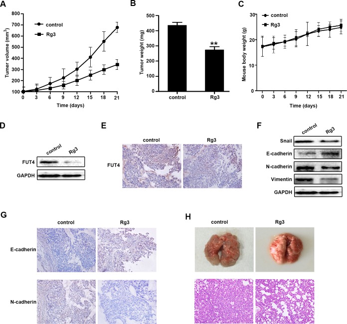
Figure 6. Ginsenoside Rg3 inhibited the growth of NSCLC xenograft tumors, EMT in vivo and tumor metastasis in tail vein injection mouse model.
A549 cells-xenografted nude mice (n = 5 per group) were injected with PBS (control) and Rg3 (10 mg/kg body weight) for 21 days. Tumor volume A., tumor weight B. and body weight C. were presented. FUT4 expression was detected by Western blot D. and immunohistochemical staining E. in xenograft tumor tissues. Snail, E-cadherin, N-cadherin and Vimentin expression were detected by Western blot F., and immunohistochemical staining of E-cadherin and N-cadherin G. in xenograft tumor tissues. GAPDH was used as an internal control (magnification, 200x). H. Pictures of mouse lung tissues (upper panel) and lung segments stained with HE (lower panel) in the control and Rg3 treatment groups after tail vein injection for 2 months. HE staining of lung segments (magnification, 200x). The statistical analysis of tumor weight is shown (**, P < 0.01). The data are presented as the mean ± SEM of three independent experiments.

