
Figure 3. SK053 covalently binds to PRDX1 in Raji cells.
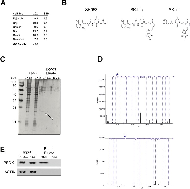
A. Cytostatic/cytotoxic effects of SK053 on human BL cell lines and normal germinal center B cells (GC B cells). BL cell lines were incubated with SK053 for 48 h and subjected to a MTT viability assay. The LC50 was calculated in Graphpad Prism 5 by nonlinear regression dose-response analysis with variable slopes. The SEM was calculated based on two independent experiments. GC B cells isolated from human tonsils (n = 3) were isolated and cultured as described in Methods. Number of viable cells after 48 h treatment with SK053 was assessed using Muse™ Cell Analyzer (Merck Millipore). LC50 was calculated in Graphpad Prism 5, as described above for BL cell lines. B. Chemical structure of SK053, its biotinylated derivative SK-bio, and the inactive biotinylated analog devoid of the electrophilic center, SK-in. C. Raji-sub cells were incubated with SK-bio or SK-in for 2 h, lysed, and biotin-labeled proteins were affinity-purified on avidin-coated beads. Total protein was resolved by SDS-PAGE and visualized by silver staining. The arrow indicates the band that was excised and identified by mass spectrometry. D. Tandem mass spectra of the Cys-173-containing peptide, HGEVCPAGWKPDGSDTIKPDVQK. The site of cysteine modification is marked with a star. The upper panel spectrum corresponds to a peptide modified with iodoacetamide (+57.021), with the parent ion m/z 802.731 and a charge 3+. The bottom panel presents the spectrum of a peptide in which cysteine bears an inhibitor (+466.225), with parent ion m/z 704.600 and a charge 4+. E. The same samples as in C. were subjected to immunobloting using antibodies specific to PRDX1 and β-actin (ACTIN).