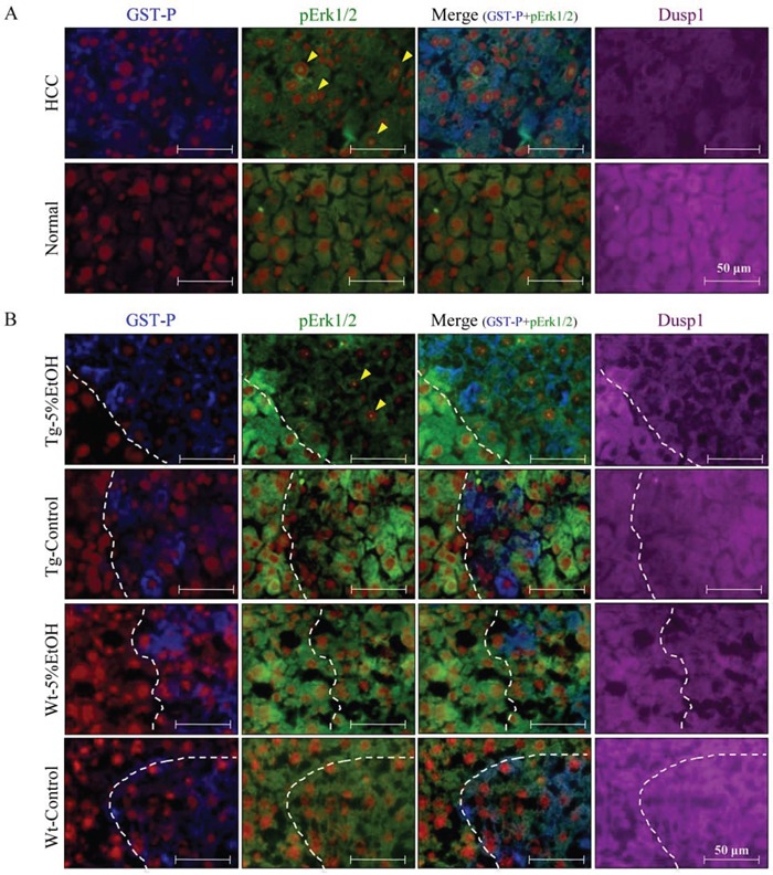
Figure 5. Localization of GST-P (blue), pErk (green), Dusp1 (purple), and nuclei (red) by triple immunofluorescence staining in HCC and normal tissue of the Tg-5%EtOH group.

A. and GST-P positive foci of each group B. Nucleolar pErk positive hepatocytes are indicated by arrowheads.
