
Figure 1. Ionizing radiation alters cellular redox and ER homeostasis in malignant gliomas.
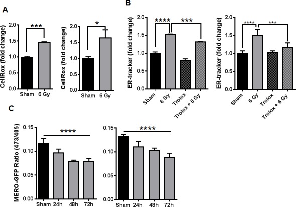
A. Measurement of total ROS by flow cytometry of cells stained with CellRox Deep Red 48h after 6 Gy IR. B. The abundance of ER-tracker staining was assayed by flow cytometry 48h after 6 Gy IR in cells pretreated with DMSO or 50uM trolox for 3h. C. The change in GFP excitation peak was measured in cells transduced with MERO-GFP to determine ER-redox status 24, 48 and 72h after irradiation, shown is the ratio of fluorescence from excitation at 473nm and 405nm. In all graphs, data shown are the Means ±SD (n = 3). *P < 0.05, ***P < 0.001, ****P < 0.0001.