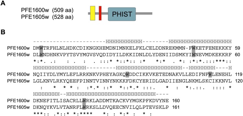
Fig 3. Protein structure of the phist genes PFE1600w and PFE1605w.
A) PFE1600w and PFE1605w possess a similar protein architecture, composed of a recessed signal peptide sequence (yellow rectangle) followed by a PEXEL/HT motif (red rectangle) and a PHIST domain (green rectangle). The numbers in the schematics indicate the lengths in aa for each protein. B) Amino acid sequence alignment of the PHIST domains from PFE1600w and PFE1605w. Tryptophan residues are shaded in gray. The predicted helical segments are shown above the alignment, marked by “H”. Below the alignment, stars indicate identical aa residues, 2 dots indicate conserved substitutions and one dot indicates semi-conserved substitutions.

