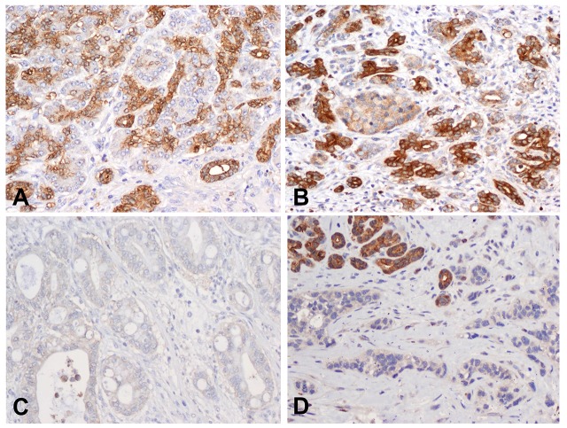
Fig 1. Expression of MAP4K5 in non-neoplastic pancreas and pancreatic ductal adenocarcinoma samples.
Representative micrographs show the expression of MAP4K5 in normal pancreatic tissue (A) and chronic pancreatitis (B). The benign pancreatic ductal cells in normal pancreas and chronic pancreatitis show strong cytoplasmic staining for MAP4K5. The adjacent pancreatic acinar cells and pancreatic islet cells are either negative or have very low expression of MAP4K5. C & D, Representative micrographs show the loss of MAP4K5 expression in two different pancreatic ductal adenocarcinoma samples. Strong cytoplasmic staining for MAP4K5 in benign pancreatic ductal cells in the left upper corner in D served as internal positive control. Original magnifications: 200X.

