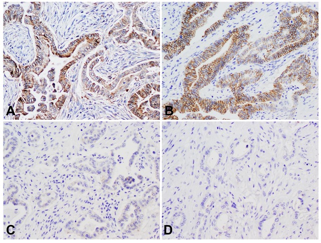
Fig 2. Expression of MAP4K5 correlates with the expression of E-cadherin in pancreatic ductal adenocarcinomas.
A & B, Representative micrographs show strong cytoplasmic staining for MAP4K5 and strong membranous staining for E-cadherin in a pancreatic ductal adenocarcinoma. C & D, Representative micrographs show loss of MAP4K5 expression and the loss of E-cadherin expression in a pancreatic ductal adenocarcinoma. Original magnifications: 200X.

