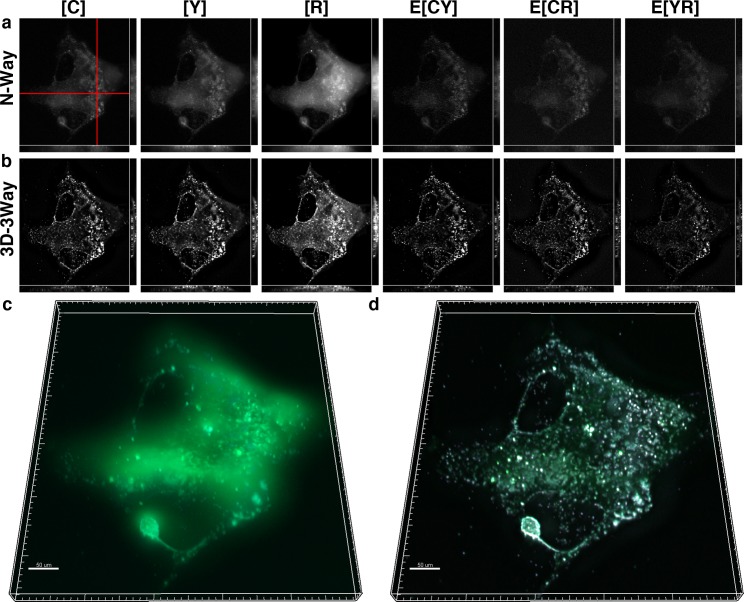
Fig 5. 3D-3Way FRET provides improved estimates for the concentration and localization of HIV-Gag oligomerization in fixed cells.
Cos7 cells expressing CFP-Gag, YFP-Gag, and RFP-Gag were fixed and imaged 30 hrs post transfection. The oligomerization of Gag into punctate structures can be seen by direct application of N-Way FRET (a). Following 20 iterations of 3D-3Way FRET (b) the FRET complexes are largely restricted to the puncta and the axial resolution is greatly improved. 3D composite rendering of [C] (cyan) [Y] (green) and E[CY] (magenta) shown following N-Way unmixing (c) and following 3D-3Way reconstruction (d). The reconstructed image highlights the improved axial resolution.

