Fig. 1.
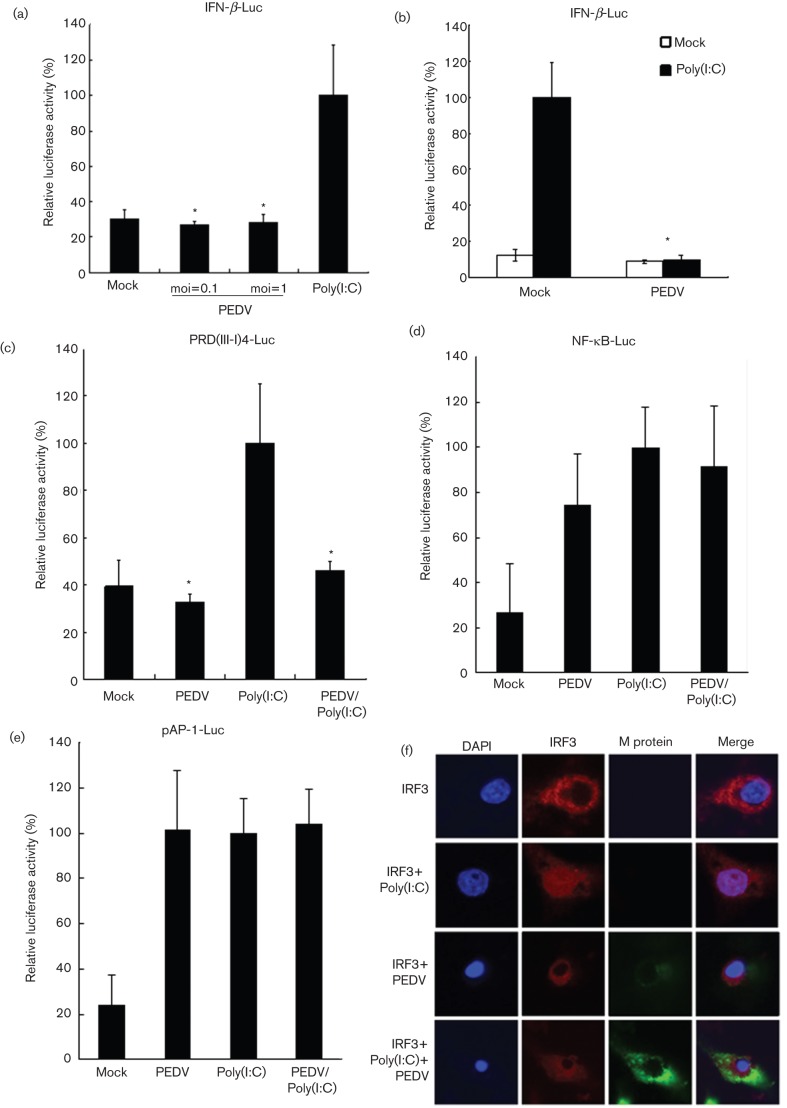
PEDV does not induce IFN-β production and blocks dsRNA-induced IFN-β promoter activation. (a) PEDV does not induce IFN-β production. Vero E6 cells were cotransfected with IFN-β-Luc and an internal control plasmid pRL-TK, followed by PEDV infection at an m.o.i. of 0.1 or 1. Poly(I : C) was used as a positive control. Cells were harvested and subjected to a Dual-luciferase assay after PEDV infection or poly(I : C) transfection. The results were expressed as mean relative luciferase (firefly luciferase activity divided by Renilla luciferase activity) with standard deviation from repeated experiments carried out in triplicate. (b) PEDV blocks dsRNA-induced IFN-β promoter activation. Vero E6 cells were infected with PEDV at an m.o.i. of 0.1. Cells were then cotransfected with IFN-β-Luc and pRL-TK. Twenty-four hours later, cells were transfected with or without poly(I : C). Cells were subjected to Dual-luciferase assay as described in (a). (c–e) PEDV inhibits dsRNA-induced activation of IRF3 but not of NF-κB and AP-1. Vero E6 cells were infected or mock-infected with PEDV at an m.o.i. of 0.1, and then cotransfected with pRL-TK and PRD(III-I)4-Luc (c), pNF-κB-Luc (d), or pAP-1-Luc (e), respectively. Twenty-four hours later, cells were transfected with or without poly(I : C). Luciferase activities were measured as described in (a). Asterisks indicate statistical significance (P<0.05). (f) PEDV inhibits dsRNA-induced IRF3 nuclear translocation. Vero E6 cells were transfected with IRF3 expression construct. After 12 h, cells were infected or mock-infected with PEDV at an m.o.i. of 0.1. Another 12 h later, cells were transfected or mock-transfected with poly(I : C). Fluorescence was examined by using a confocal microscope.

